Monday, April 11, 2016

Diagnosis: Bilateral Nephrolithiasis

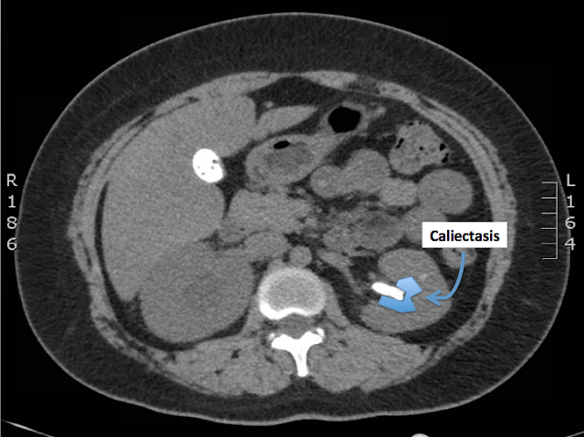
The patient's CT urogram showed bilateral nephrolithiasis with a large obstructing calculus in the right ureteropelvic junction with associated moderate hydronephrosis

There is also a large staghorn calculus in the mid-to-upper pole of the left kidney, associated with severe left upper pole caliectasis

The patient underwent interventional radiology-guided right nephrostomy tube placement without complication. Following the procedure the patient's pain improved, was able to tolerate po and ambulate. She was discharged 2 days later. 


Nephrolithiasis


Epidemiology
  • typically adults ages 20-50 years old
  • 3:1 male:female ratio
  • most stones are calcium oxalate or mixed calcium oxalate and phosphate 
  • less common: struvite (magnesium-ammonium phosphate --> urea-splitting bacteria)
    • Proteus, Klebsiella, Pseudomonas 
  • less common: uric acid (i.e. gout) 
  • 90% of stones < 5mm will pass spontaneously (> 5mm unlikely to pass) 
  • 40% of symptomatic patients will have a recurrence 
Pathogenesis
  • urine becomes supersaturated with a particular mineral 
  • poor oral intake or excessive urinary losses 
  • excess secretion of a mineral (i.e. hypercalciuria, hyperoxaluria)
  • stones form in renal collecting system and pass into ureter 
Clinical Features
  • abrupt onset of flank pain, radiates to abdomen and groin
  • nausea and vomiting are common
  • patient often unable to find a comfortable position
  • fever is atypical --> consider alternative diagnoses if present
  • abdomen usually nontender; consider abdominal aortic aneurysm if pulsatile mass palpated or bruit auscultated
  • costovertebral angle tenderness is inconsistent
Urinanalysis 
  • hematuria usually present, about 15% however will not have hematuria 
  • WBCs and bacteria should raise concern for infection 
  • pH >7.6 = concern for struvite and urea-splitting bacteria 
Blood work
  • CBC and electrolytes typically are normal
  • BUN and Creatinine are usually normal even in context of obstructive uropathy if other kidney is functioning
  • Check calcium 
**Rule out vascular catastrophe! (i.e. AAA, iliac aneurysm, etc)** 

Imaging
  • CT urogram = gold standard 
    • sensitive and specific, can measure degree of obstruction if present, may detect alternative diagnoses, no IV contrast needed
  • US
    • great for detecting and measuring renal obstruction, can detect stones in the kidneys, often misses stones in the ureters
Management
  • analgesia with NSAIDs (first-line), opioids, or both
  • antiemetics 
  • IV hydration 
  • alpha-blocking agents such as Tamulosin to aid stone passage is controversial (currently recommended by the American Urological Association) 
  • absolute indications for admission:
    • intractable nausea and vomiting, or intractable pain 
    • obstruction with infection
  • relative indications for admission:
    • stone >5 mm
    • high-grade obstruction
    • solitary kidney
    • deteriorating renal function 


References

Lopez BL, Brooks M, Emergency Medicine: A Focused Review of the Core Curriculum, 1st Ed, 2008, Chapter 17: Renal and Urological Disorders, pages 989-994

Discite Exerceo Adservio


Saturday, April 2, 2016

Flank Pain

A 34 year-old female presents to the emergency department with 1 day of worsening right-sided flank and lower back pain. The pain starts in her right lower back and right flank and radiates to her stomach. She describes the pain as sharp, 10/10, constant, improved by intravenous morphine in the emergency department to 4/10. The pain is not made worse by by urination, food, or movement. She denies having any recent fever, chills, dysuria, or hematuria. She denies any vaginal bleeding. 

A CT urogram was obtained and is shown below:


What is the diagnosis?

How would you manage this patient?

Share your thoughts in the comments section below.

The diagnosis for this case will be posted in approximately 1 week.

Sunday, December 6, 2015

Diagnosis: Acute Pericarditis

Review of the patient's ECG demonstrated diffuse ST-elevations and questionable PR-depression consistent with acute pericarditis. His chest x-ray was interpreted as no acute cardiopulmonary disease. 

The patient's chest exam revealed a faint friction rub. Bedside cardiac ultrasound revealed a mild-to-moderate pericardial effusion. 

The patient's troponin came back at 0.29 and returned to undetectable levels on serial checks. A subsequent formal ECHO study revealed normal cardiac function including normal ejection fraction. 

The patient was admitted to the hospital overnight. His pain improved with oral ibuprofen. A repeat ECG the next day showed resolution of the original ST-elevations and the patient was discharged home with follow-up with his primary care provider. 


Acute Pericarditis

Normally 15-60 cc of physiological fluid between visceral and parietal layers of the pericardium

Inflammatory conditions increase the amount of fluid in this space --> can result in cardiac tamponade depending on the amount of additional fluid and rate at which it increases

Etiologies: viral, idiopathic, post-myocardial (Dresser's syndrome), neoplastic, infectious, uremic, radiation, connective tissue disorders (SLE, RA, Sjogren's syndrome, etc)  

Patient history
  • sharp pain of prolonged duration (often days) 
  • pleuritic component
  • worse when supine
  • improved when sitting forward
  • prodrome of fever and malaise, not always present but common 
Clinical findings = pericardial friction rub (best heard over left lower sternal border) 

ECG findings
  • Stage 1 - diffuse ST-elevations with concave-upwards contour, PR-depressions (most common in II, aVF, V4-6)
  • Stage 2 - ST segments become isoelectric and T waves flatten
  • Stage 3 - symmetric T wave inversion throughout ECG 
  • Stage 4 - normalization 

**PEARL** 
Anything that irritates the outer epicardium of the myocardium will produce ST-elevations. This explains why only transmural infarcts involving the epicardium result in ST-elevation, and why ST-elevations indicate that your pericardiocentesis needle is making contact with the outer myocardium.

Management
  • evaluate and treat underlying cause (e.g. dialysis for uremia)
  • NSAIDs 


References

Colletti JE, Tabas JA, Emergency Medicine: A Focused Review of the Core Curriculum, 1st Ed, 2008, Chapter 2: Cardiovascular Disorders, pages 237-242


Discite Exerceo Adservio


Monday, November 30, 2015

A Little Post-Cold Chest Congestion

A 22 year-old male with history of ventricular septal defect repaired at 2 years of age, and gastritis presents to the emergency department with a chief complaint of chest pain. 

He endorses having a sore throat and other cold symptoms for the past few days that have mostly resolved by now. He reports taking some DayQuil earlier today and later drank a 24 ounce beer. 

A few hours later he started to experience some substernal chest pain described as a constant pressure and occasional burning sensation with mild sweating and shortness of breath. The pain is not relieved by resting or leaning forward. He denies have any associated nausea, vomiting, reflux symptoms, or recent trauma to his chest. 


An ECG  and chest x-ray were obtained and are shown below: 


What is the diagnosis?

How would you manage this patient?

Share your thoughts in the comments section below.

The diagnosis for this case will be posted in approximately 1 week. 

Sunday, October 11, 2015

Diagnosis: Spontaneous Pneumothorax

The patient's chest x-ray revealed a right apical pneumothorax estimated 10-15% in size with some mild right basilar atelectasis. No focal consolidation or edema. No acute osseous abnormality.

The pulmonology service was consulted and recommended a period of observation on 100% oxygen. A repeat chest x-ray after 6 hours showed no significant change in size. At discharge the patient continued to endorse pain with deep inspiration, but otherwise reported feeling comfortable. She was given a follow-up appointment with pulmonology for repeat imaging in a week or two.


Pneumothorax


Air between the visceral and parietal pleura

Due to an alveolar-pleural defect between intrapleural space (normally under negative pressure) and the airways (positive pressure)

Defect leads to equalization of pressure until the defect is closed


Simple
  • lung collapse on the side of the defect --> decreased lung capacity
  • VQ mismatch --> potential hypoxemia depending on size and comorbidities
Tension
  • defect acts as a one-way valve --> accumulation of air in the intrapleural space
  • mediastinal contents shift away from affected side --> compresses opposite lung
  • hypoxia and impaired venous return --> cardiovascular collapse and death
Causes
  • Traumatic - blunt or penetrating trauma 
  • Iatrogenic - often due to penetrating trauma (i.e. procedure) 
  • Spontaneous - no clear precipitating cause
    • Primary (no lung disease) = 2/3 cases
      • Risk factors: male, smoker, tall, change in ambient pressure, Marfan's
    • Secondary (history of lung disease, usually COPD) 
      • Risk factors: asthma, cystic fibrosis, lung abscess, TB, interstitial lung disease, malignancy, PJP
Presentation
  • sudden onset unilateral chest pain, usually pleuritic, dyspnea, cough
  • tachycardia, hyperresonance to percussion, diminished breath sounds
Diagnosis
  • clinical suspicion should prompt imaging
  • CXR - classically shows pleural line parallel to contour of chest wall with gap of absent lung markings
  • CT chest = very sensative
  • Grade: small, moderate, large, total
Management
  • largely depends on the clinical status of the patient and the size of the pneumothorax
  • observation (6 hours) 
    • small size pneumothorax <20%
    • otherwise young healthy patient
    • intrinsic reabsorption 1%-2% per day (increases to 4%-8% on 100% O2)
    • may discharge after observation if reliable follow up established for repeat CXR
  • simple catheter aspiration 
    • moderate size pneumothorax >20%
    • less invasive but not always successful
  • tube thoracostomy
    • most common therapy, especially when more conservative measures fail
    • treatment of choice with pleural effusion, respiratory distress, tension pneumothorax
    • 20-28 French standard, >28 preferred if pleural fluid present
    • water-seal valve (Heimlich or flutter valves) don't require hospitalization
    • complications: malposition, infection, pain, obstruction, reexpansion pulmonary edema
management of tension pneumothorax to be discussed in a future post


References

Levy SJ, Lex JR, Emergency Medicine: A Focused Review of the Core Curriculum, 1st Ed, 2008, Chapter 4: Thoracic and Respiratory Disorders, pages 237-242


Discite Exerceo Adservio

Sunday, October 4, 2015

My Shoulder Really Hurts...

A 27 year-old female presents to the emergency department with chief complaint of right-sided shoulder pain radiating to the chest that started 3 days ago. 

She reports that the pain was initially very severe and exacerbated by every breath on the first day of symptoms. The pain began after waking that morning. The patient went to a local urgent care clinic where she had a shoulder x-ray done that did not show a fracture or dislocation and was discharged with the diagnosis of impingement syndrome. The pain has improved somewhat over the last 2 days but has continues to experience pain with deep inspiration. The pain is also made worse by lying flat.

She denies any recent trauma to her shoulder or chest. She also denies any history of recent long car or plane rides, OCP use, recent surgery, or prior deep venous thrombosis. 

She denies having any shortness of breath, fevers, chills, nausea or vomiting.

Vital signs are within normal limits. The patient appears resting comfortably in bed.

A 2-view chest x-ray was obtained and is shown below: 


What is the diagnosis?

How would you manage this patient? 

Share your thoughts in the comments section below.  

The diagnosis for this case will be posted in approximately 1 week.

Monday, August 17, 2015

Diagnosis: Pancreatic Adenocarcinoma

CT of the abdomen and pelvis revealed a mass within the pancreatic head and uncinate process, most likely representing pancreatic neoplasm. There was mass effect noted with resultant mild intrahepatic and extrahepatic biliary duct and pancreatic duct dilatation. Also evidence of pancreatitis likely secondary to pancreatic tumor.

The patient's lipase level returned >2400

A subsequent endoscopic retrograde cholangiopancreatography (ERCP) revealed a dilated common bile duct and pancreatic duct but no significant intrahepatic biliary dilation.  With ultrasound a hypoechoic 24 X 23 mm mass was seen in the head of pancreas with upstream dilation of both ducts. Vessels looked clear of the mass. Small amount of ascites noted. Small peritumor lymph node seen.  

Preliminary diagnosis by fine needle aspiration of the mass suggested neoplasm.

Final biopsy results: pancreatic adenocarcinoma.

The patient was discharged on hospital day 3 with follow up with gastroenterology and oncology.


Pancreatic Cancer


Background 
  • 4th most common cause of cancer-related death in the United States
  • Ductal adenocarcinomas account for 95% of malignant pancreatic tumors
  • Pancreatic head involved in 70% of cases
  • Especially lethal --> 5-year survival rate < 5%
  • Few early symptoms --> most patients diagnosed late 
Risk Factors
  • smoking
  • advanced age
  • high fat diet
  • positive family history
Presentation
  • constant dull pain in the epigastrium
  • weight loss (more due to anorexia than malabsorption) 
  • painless jaundice (75% of patients) 
  • enlarged palpable painless gallbladder = Courvoisier's sign
  • glucose intolerance progressing to diabetes
  • migratory inflammatory thrombophlebitis = Trousseau's sign of malignancy
Diagnosis = CT 

Management
  • complete resection = only effective treatment
  • few tumors (<20%) diagnosed early enough for curative surgical therapy
  • complications presenting to the ED
    • bowel obstruction
    • jaundice
    • problems with pain control --> do not withhold narcotics
  • consult oncology to address end-of-life issues


References

Hemphill RR, Santen SA, Rosen's Emergency Medicine Concepts and Clinical Practice, 8th Ed, 2014, Chapter 91: Disorders of the Pancreas


Discite Exerceo Adservio